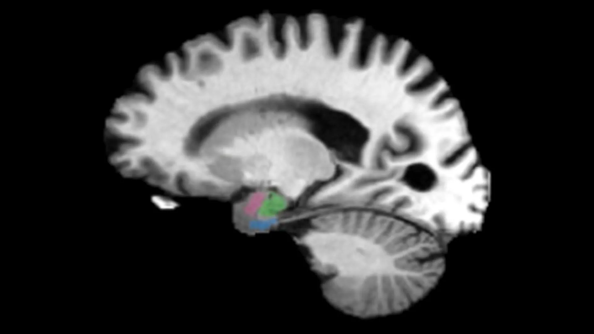
Johns Hopkins University researchers have identified two brain regions that show tissue loss earlier than the hippocampus in Alzheimer's disease, potentially offering a way to detect the condition years before memory problems develop.
By examining hundreds of MRI and PET scans from two independent studies, the team found that the entorhinal cortex and amygdala begin shrinking up to a decade before cognitive symptoms appear. The entorhinal cortex helps retrieve stored memories, while the amygdala processes emotional memory.
"We have found that there are early changes in the brain manifesting the clinical biomarkers of atrophy as much as a decade before there are clinical symptoms," said Michael Miller, biomedical engineer and senior author. "It's at least a 5-to-10-year play where your brain is changing."
The researchers, including Marilyn Albert, director of the Johns Hopkins Alzheimer's Disease Research Center, observed consistent patterns in both study groups across all disease stages. The tissue loss in these two regions correlated with tau protein accumulation, a hallmark of Alzheimer's pathology. Tau normally helps maintain nerve cell structure but can form toxic tangles in Alzheimer's.
The findings appeared in NeuroImage Reports and were presented at Neuroscience 2026, the annual meeting of the Society for Neuroscience.
The team plans future research to identify molecular changes surrounding tau accumulation in these regions. Miller stated, "Once we know more about the molecular neighborhoods, we can develop therapies." The researchers note that additional work is necessary before these brain changes become a reliable diagnostic tool in clinical practice.