# Dinosaur's Healing Rib Reveals 66-Million-Year-Old Biology Through Advanced Imaging
Scotty, the world's largest known Tyrannosaurus rex skeleton, carried a broken rib that preserved something paleontologists rarely encounter: the internal architecture of a dinosaur's healing process frozen in stone for 66 million years.
Researchers used neutron and X-ray imaging to peer inside the damaged bone without destroying it, uncovering an intricate network of blood vessels that formed as the massive predator recovered from injury. The fossilized vascular system offers an unusually detailed window into how T. rex physiology responded to trauma during the Late Cretaceous period.
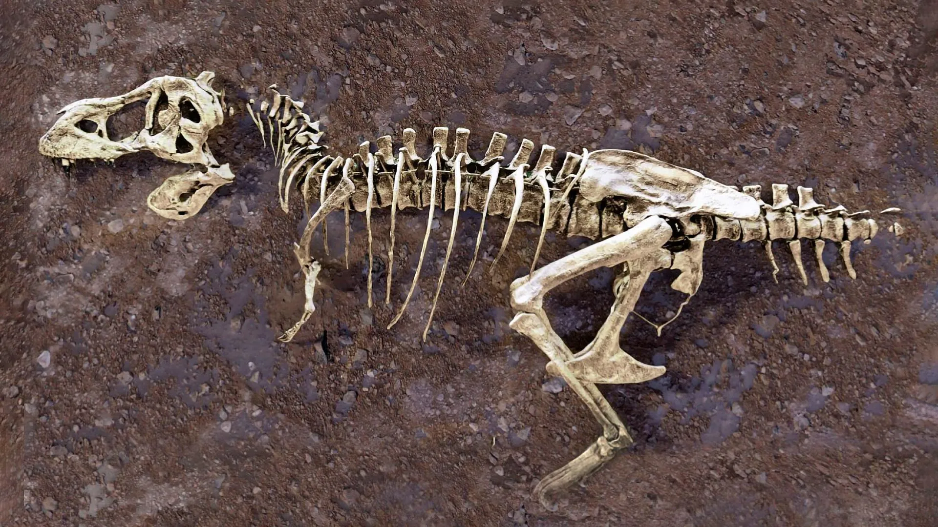
Scotty, discovered in Saskatchewan, Canada, and housed at the Royal Saskatchewan Museum, stands as the most complete and heaviest T. rex skeleton ever found. The dinosaur measures over 13 meters in length and weighs an estimated 11,600 kilograms. Scientists estimate this individual lived roughly 66 million years ago, near the end of the Cretaceous era. The specimen bears evidence of multiple injuries and survival, suggesting a long and combat-heavy life.
The broken rib represents one of several wounds visible on Scotty's skeleton. What makes this particular fracture remarkable is how completely the healing process preserves internal bone remodeling. When bones break and heal, blood vessels proliferate to deliver nutrients and remove damaged tissue. These vessels typically decompose and leave no trace in the fossil record. This specimen captured them in mineralized form.
Neutron imaging proved especially valuable for this work. Unlike conventional X-rays, neutron beams penetrate heavy elements and interact differently with various materials. This capability allowed researchers to visualize the three-dimensional pathways of fossilized blood vessels running through the bone tissue. X-ray imaging complemented the neutron data, providing additional structural detail from different angles.
The team did not release their names or institutional affiliations in the available source material, limiting credit attribution. However, the research represents collaboration among multiple institutions with expertise in paleontology, medical imaging, and neutron physics. Such interdisciplinary approaches have become standard for studying dinosaur biology in recent years.
The findings extend beyond simple bone healing mechanics. The vascular network provides information about metabolic rates, growth patterns, and tissue repair efficiency in large theropod dinosaurs. Understanding how Scotty's body responded to injury offers insights into T. rex physiology during adulthood. The data suggests these animals possessed sophisticated biological systems capable of managing serious trauma and recovering successfully.
Scotty's survival despite multiple injuries raises questions about T. rex behavior and ecology. The broken rib and other wounds might have resulted from predator-prey interactions, intraspecific combat between T. rex individuals, or environmental hazards. Each scenario carries different implications for understanding social structure and hunting strategies in Late Cretaceous ecosystems.
Non-destructive imaging technologies continue transforming paleontology. Researchers can now examine irreplaceable fossils at scales previously requiring physical sectioning that destroyed the specimens. This shift has revealed anatomical details in specimens worldwide. Future scanning of other healed injuries in Scotty and comparable fossils could generate datasets tracking injury frequency, healing success rates, and disease patterns across theropod populations.
Scotty's broken rib demonstrates how cutting-edge physics applied to ancient biology yields discoveries that reshape understanding of extinct animals. The preserved blood vessels represent not just anatomical curiosity but evidence of metabolic capacity, survival instinct, and biological resilience in one of Earth's most formidable predators.