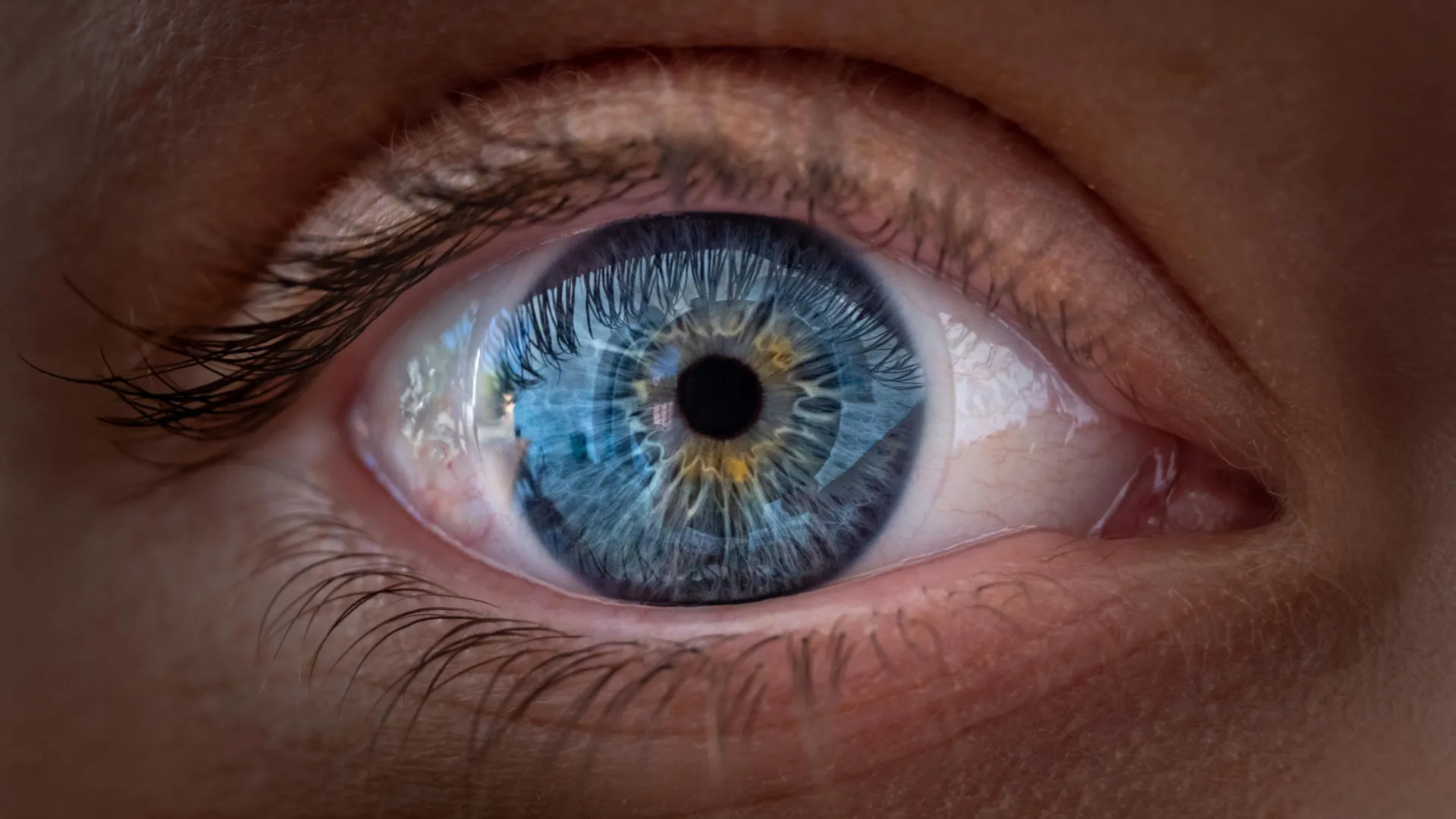
Scientists at Johns Hopkins University discovered that vitamin A and thyroid hormones work together to reshape cone cells in the developing human retina before birth. The research, published in Proceedings of the National Academy of Sciences, shows that blue light-detecting cells in the foveola (the central retina region) transform into red and green light-detecting cells during weeks 10 through 14 of fetal development.
Robert J. Johnston Jr., an associate professor of biology at Johns Hopkins who led the study, explained the mechanism. "First, retinoic acid helps set the pattern. Then, thyroid hormone plays a role in converting the leftover cells," Johnston said. "That's very important because if you have those blue cones in there, you don't see as well."
The findings challenge a 30-year-old model suggesting that blue cones simply migrated away from the foveola. Johnston stated, "These cells actually convert over time, which is really surprising."
To make this discovery, Johnston's team grew miniature human retinal tissues called organoids from fetal cells. These laboratory-grown tissues allowed researchers to observe developmental changes that would be impossible to study during pregnancy. The foveola, though occupying only a small fraction of the retina, accounts for approximately 50 percent of human visual perception, according to the researchers.
The study focuses on retinoic acid, a molecule derived from vitamin A that regulates cell development. However, Johnston noted that the discovery "does not establish that taking vitamin A supplements can improve sharp vision in adults."
The researchers believe the findings could advance treatments for macular degeneration, glaucoma, and other vision-loss conditions. Johnston's team is now working to develop retinal organoids that could eventually produce healthy light-detecting cells for transplantation into damaged retinas.