# Brain Textbooks Had It Wrong: Axons Aren't Smooth Tubes
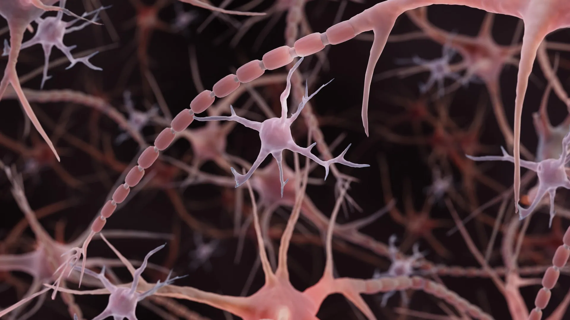
For over 100 years, every neuroscience textbook depicted axons as uniform, smooth tubes transmitting electrical signals from one neuron to the next. New research reveals this foundational drawing was incorrect. Axons actually display a pearl-like structure along their length, with regular constrictions that resemble beads on a string rather than the featureless cylinders shown in standard diagrams.
A team of neuroscientists discovered these structures, called nodes of Ranvier on a more granular scale, function as dynamic regulators of signal transmission. The pearl-like geometry appears throughout healthy axons and responds to neural activity, suggesting the brain may have evolved this architecture to fine-tune electrical communication speed.
The discovery challenges a core assumption embedded in neurobiology education and research for generations. When Santiago Ramón y Cajal first mapped neurons in the late 1800s using his revolutionary staining techniques, his hand-drawn illustrations established the smooth axon model that persisted unchallenged in published materials. That representation became canonical, reproduced in countless textbooks without revisiting whether it accurately reflected reality.
The constricted, pearl-like appearance of axons appears to be a natural feature present across multiple neuron types and species. These structures can enlarge or contract in response to neural firing patterns and metabolic demands. The dynamic nature of these axonal pearls suggests they serve a biological function rather than representing an artifact of tissue preparation or microscopy.
This geometry may operate as a biological switch for controlling conduction velocity. When axons fire repeatedly, the pearls can change diameter, potentially altering how quickly action potentials propagate down the axon. This mechanism could allow neurons to adjust communication speed based on behavioral demands or learning requirements, adding another layer of neural plasticity.
The research implications extend beyond textbook corrections. Understanding axonal architecture at this scale could reshape how scientists interpret neural recordings and computational models of brain function. Many existing simulations of neural circuits treat axons as uniform conductors, an assumption now known to be incomplete. Future models incorporating pearl-like geometry might predict neural behavior with greater accuracy.
The discovery also suggests new targets for understanding neurological diseases. Conditions like multiple sclerosis, where myelin sheaths deteriorate, and neurodegenerative diseases showing axonal degeneration might involve disruption of these pearl-like structures. Examining whether disease progression correlates with changes in axonal geometry could open diagnostic or therapeutic approaches.
This work exemplifies how foundational knowledge in science can persist without scrutiny. The smooth axon model became so embedded in the field that researchers rarely questioned whether textbook diagrams matched actual biological structures. Modern imaging techniques finally allowed scientists to examine axons at sufficient resolution to detect the pearl-like morphology.
The findings underscore the importance of revisiting established models with contemporary tools. What appeared settled for a century turned out incomplete. Future neuroscientists will study axon structure with more precise understanding, potentially uncovering additional features that current imaging technology cannot yet resolve. The smooth tube of yesteryear's textbooks now belongs to scientific history.