# How Glucose and Signals Guide the Brain's Blueprint Before Birth
UCLA researchers have identified two fundamental mechanisms that orchestrate the formation of the human cerebral cortex during fetal development. The work, which focuses on radial glia—the stem cells that generate most of the cerebral cortex—reveals how metabolic activity and molecular signals work together to direct neural development.
Radial glia cells do not behave uniformly. Instead, their behavior shifts based on how they metabolize glucose, the primary fuel source for developing neural tissue. This metabolic switch acts like a dimmer controlling which developmental pathways activate in these stem cells. The UCLA team discovered that glucose processing influences whether radial glia remain as self-renewing stem cells or differentiate into specialized neurons.
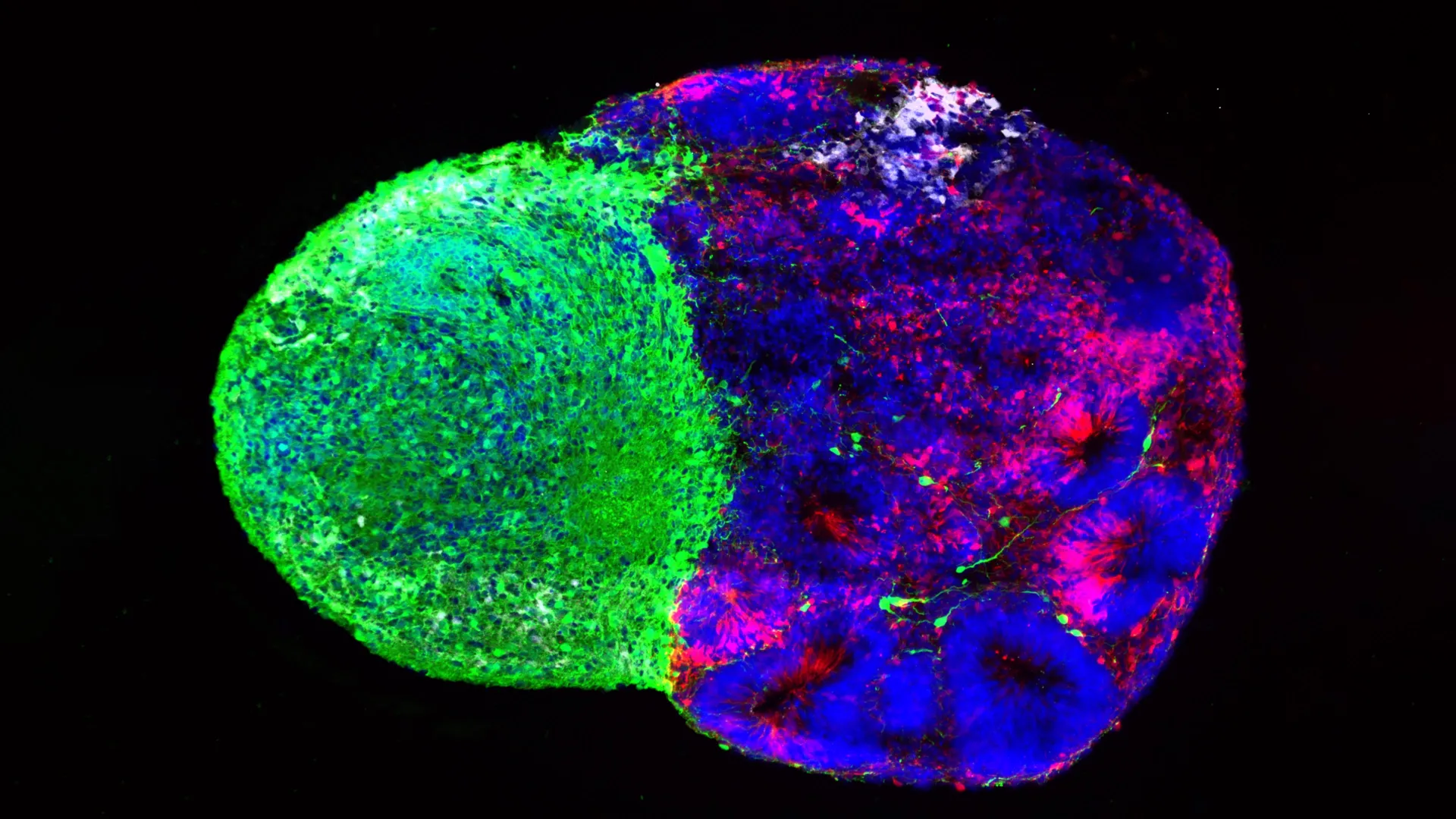
The second layer of control involves physical signals from the thalamus, a region deep in the brain that serves as the brain's central relay station. Thalamic neurons send long-range connections that contact radial glia cells during development. These connections deliver biochemical signals that alter the cell fate decisions of radial glia, changing which types of neurons they produce.
The combination of these two influences proved particularly important for generating upper-layer cortical neurons. These neurons form the superficial layers of the cerebral cortex and play critical roles in higher cognitive functions like consciousness and complex reasoning. Upper-layer neurons are proportionally larger and more numerous in humans than in other primates or rodents, making them a key feature of human brain evolution.
The research addresses a longstanding puzzle in neuroscience: how does the developing brain ensure the correct proportions of different neuron types form at the right times and places? Previous studies showed that radial glia respond to various environmental cues, but the relative importance of metabolic versus signaling mechanisms remained unclear.
UCLA researchers used cultured neural tissue systems and experimental manipulations to isolate each factor's contribution. By altering glucose availability and blocking thalamic signals separately, they measured how each change affected the types of neurons produced. The results showed both factors operate together, rather than independently, to fine-tune neural development.
Understanding these mechanisms has implications beyond basic science. Disruptions in glucose metabolism or thalamic development are implicated in several neurodevelopmental conditions. Certain genetic forms of autism spectrum disorder involve mutations affecting metabolic enzymes or thalamic circuit formation. Schizophrenia research has identified abnormalities in thalamic connectivity and cortical layer formation. Mapping exactly how these systems normally function provides a framework for understanding what goes wrong in disease states.
The findings also inform efforts to generate human neurons from stem cells for disease modeling and therapeutic applications. Researchers developing replacement neurons for brain injury or neurodegeneration need to control which neuron types their stem cells produce. Knowing that glucose metabolism and thalamic-like signals influence these decisions offers new tools for directing stem cell differentiation in the lab.
The UCLA work represents incremental progress on a complex problem. The developing brain contains billions of cells coordinating development through hundreds of signaling pathways. This research isolates two important ones, but many other factors undoubtedly contribute to cortical formation. Future work will explore how these metabolic and signaling mechanisms interact with other developmental cues.