# Smart Nanoparticles Target Hidden Brain Cancer After Surgery
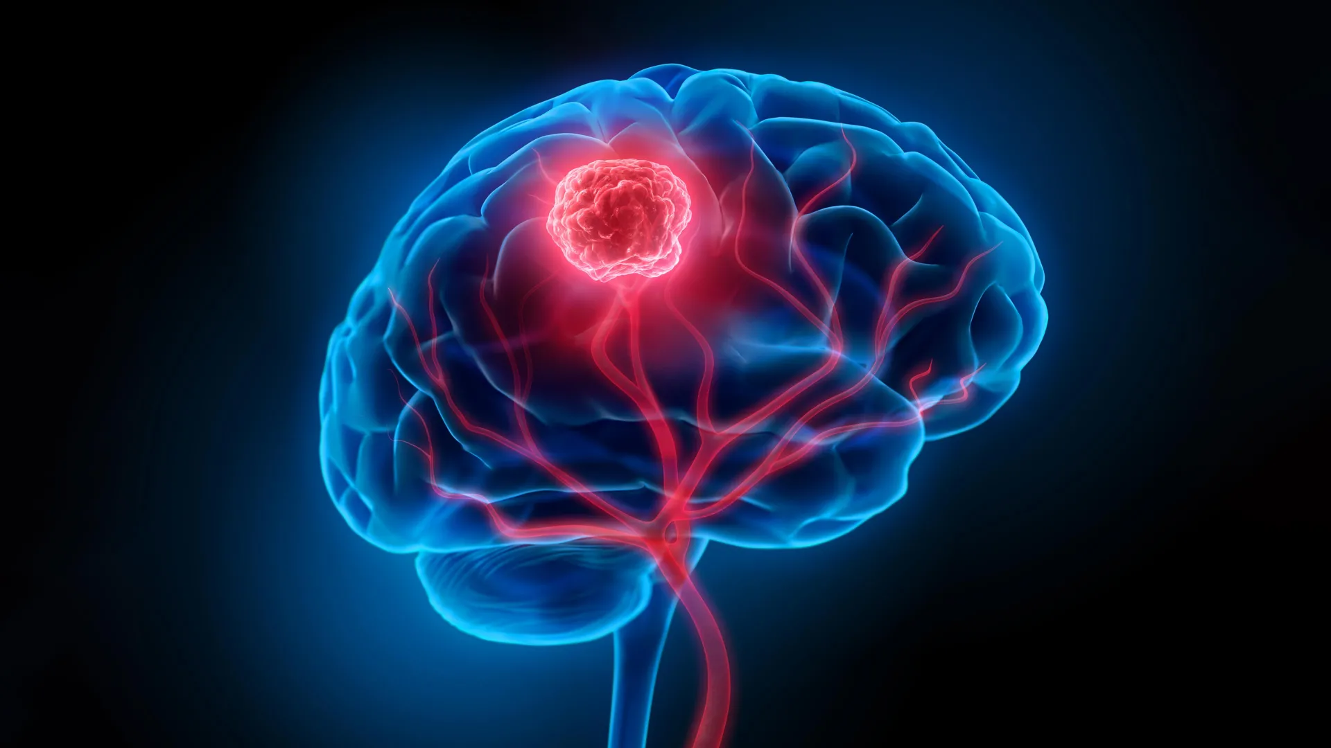
Researchers have engineered nanoparticles that perform dual roles in treating glioblastoma, the most aggressive form of brain cancer. The particles light up invisible cancer cells during surgery while simultaneously priming them for destruction afterward.
Glioblastoma kills most patients within two years of diagnosis. Surgery removes visible tumors, but microscopic cancer cells often escape detection and regrow. Current treatments like chemotherapy and radiation fail because they cannot selectively target residual disease while sparing healthy brain tissue.
The nanoparticles work through a two-stage mechanism. First, they accumulate in tumor cells and fluoresce under near-infrared light, allowing surgeons to see and remove cancer that appears normal to the naked eye. Second, the particles remain in surviving cancer cells and respond to light exposure by generating reactive oxygen species. These toxic molecules destroy the cells from within.
Researchers conducted the study in mice bearing human glioblastoma tumors. Animals treated with the nanoparticle platform combined with near-infrared light therapy showed 100% survival at 60 days post-treatment, compared to rapid death in untreated controls. More importantly, the approach prevented tumor recurrence in surviving mice, addressing the central clinical problem that makes glioblastoma so deadly.
The nanoparticles consist of a core material loaded with photosensitive dyes, surrounded by a protein coating that helps them accumulate selectively in cancer cells rather than healthy brain tissue. This selectivity reduces damage to normal neurons and glial cells. The particles measure roughly 100 nanometers, allowing them to navigate the brain's unique vascular environment.
The study emerged from interdisciplinary collaboration combining materials science, oncology, and biomedical engineering. Researchers optimized particle composition and loading strategies to maximize fluorescence intensity and reactive oxygen production while maintaining safety profiles acceptable for brain tissue. They also tested different light wavelengths to identify the most effective activation parameters.
Several limitations temper enthusiasm for immediate human trials. Mouse brains differ fundamentally from human brains in size, complexity, and blood-brain barrier function. The particles require near-infrared light penetration to activate, which presents technical challenges in deep brain regions. Surgical delivery requires developing methods to administer particles directly to the tumor bed while avoiding systemic toxicity. Long-term safety remains uncharacterized in larger animal models.
The research team must now pursue preclinical validation in larger animals and optimize the surgical procedure for clinical translation. They need to establish safe dosing windows, confirm the particles clear from the brain within acceptable timeframes, and develop imaging systems compatible with operating room equipment.
Glioblastoma patients currently experience median survival of 15 months with standard care. Any approach that prevents recurrence could extend survival substantially. The nanoparticle platform addresses a genuine unmet need, but substantial development separates promising mouse results from effective human treatment. Clinical trials would require FDA approval and careful patient selection to justify the experimental nature of the approach.