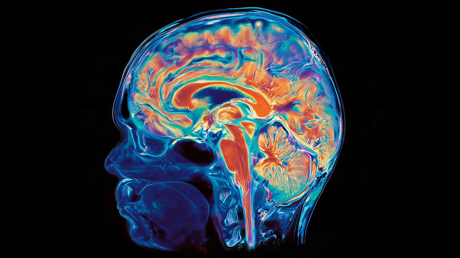
Researchers at Stanford University have uncovered evidence that the human brain may represent a fusion of two fundamentally different organs, each derived from separate evolutionary lineages. This finding reshapes our understanding of how the brain developed and offers a new avenue for studying neurodegenerative diseases.
The Stanford team identified that the brain develops from two distinct cellular systems with separate origins and specialized functions. Rather than emerging as a single unified structure, the human brain appears to have evolved when two ancient nervous systems merged over millions of years. One system likely controlled basic survival functions like breathing and heartbeat regulation, while the other handled more complex cognitive tasks. This dual-origin hypothesis explains longstanding puzzles about brain organization and suggests that what we call the brain today represents an evolutionary compromise between these two systems.
The research hinges on tracking how different neural cell populations arise during human brain development. By examining gene expression patterns and cellular lineages in developing embryonic tissue, the Stanford scientists mapped out the separate developmental trajectories that eventually integrate into a single functioning organ. This level of detail required advanced molecular techniques to trace cells back to their developmental origins.
The practical implications extend beyond evolutionary theory. The team successfully grew human hindbrain neurons in laboratory culture, a breakthrough that opens experimental possibilities previously unavailable to researchers. The hindbrain, which sits at the base of the skull and controls vital functions, commonly degenerates in several serious diseases. Amyotrophic lateral sclerosis, or ALS, progressively destroys motor neurons in the brainstem and spinal cord, paralyzing patients while leaving their minds intact. Spinal muscular atrophy, or SMA, causes similar devastation by affecting motor neuron development and survival. Other brainstem disorders like progressive supranuclear palsy and multiple system atrophy also involve hindbrain dysfunction.
Growing human hindbrain neurons in culture allows scientists to study disease mechanisms in detail without relying solely on animal models or post-mortem tissue. Researchers can expose these cells to potential drug candidates, test genetic modifications, and observe how disease processes unfold at the cellular level. This platform accelerates the discovery process and helps researchers understand why certain neurons become vulnerable in each disease.
The discovery also prompts reconsideration of how different brain regions communicate. If the brain truly consists of two integrated systems with distinct evolutionary histories, this raises questions about how they coordinate activity and share information. Understanding these integration mechanisms could illuminate why damage in one region often affects distant areas, and why some treatments targeting one system have unexpected effects elsewhere.
The Stanford findings appear positioned to reshape neurobiology textbooks and guide future research into brainstem diseases. The next steps likely involve using the lab-grown hindbrain neurons to screen drug libraries, test patient-derived genetic variants, and model disease progression in real time. This cellular platform represents a tangible bridge between evolutionary insights and therapeutic development.