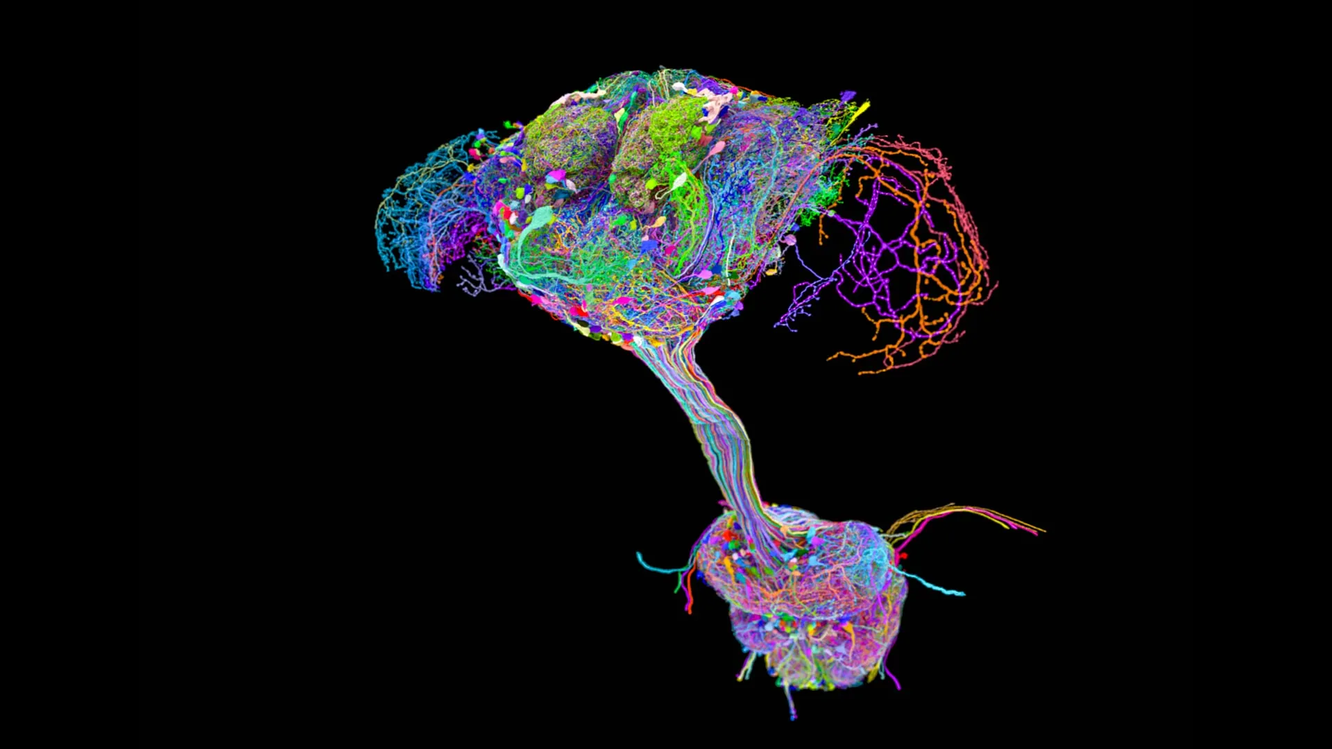
Researchers have completed a full connectome of the fruit fly central nervous system, mapping all 139,000 neural connections with extraordinary precision. This represents the first complete wiring diagram of any adult animal brain, advancing neuroscience beyond the earlier C. elegans connectome which mapped only 302 neurons in a larval worm.
The study reveals that complex behaviors like flight, navigation, and courtship arise from distributed networks of local circuits rather than hierarchical control from a central command center. Each region of the fly brain operates semi-autonomously, coordinating with neighboring circuits to produce coordinated action. This architecture challenges traditional neuroscience models that emphasize top-down control.
The connectome identifies millions of synaptic connections across approximately 3,000 neurons in the fly's brain and ventral nerve cord. Researchers used serial-section electron microscopy to image thin slices of tissue, then reconstructed connections computationally. The work involved teams at multiple institutions analyzing years of imaging data.
The findings have immediate applications for understanding human neurology. The fruit fly shares fundamental neural principles with mammals, including similar neurotransmitter systems and circuit motifs. Researchers expect the connectome will accelerate studies of learning, memory formation, and sensorimotor integration.
Key surprises emerged about circuit organization. The team discovered unexpected feedback loops between sensory and motor regions, suggesting the brain continuously updates motor commands based on real-time feedback rather than executing pre-planned sequences. They also found that neurons involved in different functions cluster together physically, contradicting assumptions about strictly segregated brain regions.
Limitations remain. A connectome provides anatomy but not function. Researchers cannot yet determine which synaptic connections are excitatory or inhibitory simply from structure. Electron microscopy also captures static anatomy, not dynamic neural activity. Mapping the complete connectome required years of work and specialized expertise, making this approach